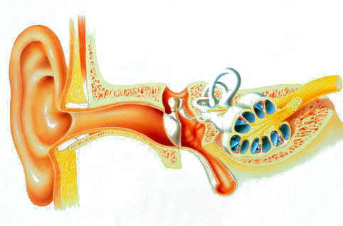
The structure of the human ear anatomy. The structure of the organ of hearing
The human ear is a unique, rather complex organ in its structure. But, at the same time, the method of its work is very simple. The organ of hearing receives sound signals, amplifies them and converts them from ordinary mechanical vibrations into electrical nerve impulses. The anatomy of the ear is represented by many complex constituent elements, the study of which is singled out as a whole science.
Everyone knows that the ears are a paired organ located in the region of the temporal part of the human skull. But, a person cannot see the device of the ear in full, since the auditory canal is located quite deep. Only the auricles are visible. The human ear is capable of perceiving sound waves up to 20 meters long, or 20,000 mechanical vibrations per unit time.
The organ of hearing is responsible for the ability to hear in the human body. In order for this task to be performed in accordance with the original purpose, the following anatomical components exist:
human ear
- The outer ear, presented in the form of an auricle and auditory canal;
- The middle ear, consisting of the tympanic membrane, a small cavity of the middle ear, the ossicular system, and the Eustachian tube;
- The inner ear, formed from a transducer of mechanical sounds and electrical nerve impulses - snails, as well as systems of labyrinths (regulators of balance and position of the human body in space).
Also, the anatomy of the ear is represented by the following structural elements of the auricle: curl, antihelix, tragus, antitragus, earlobe. The clinical auricle is physiologically attached to the temple by special muscles called rudimentary.
Such a structure of the hearing organ has the influence of external negative factors, as well as the formation of hematomas, inflammatory processes, etc. Ear pathologies include congenital diseases that are characterized by underdevelopment of the auricle (microtia).

outer ear
The clinical form of the ear consists of the outer and middle sections, as well as the inner part. All these anatomical components of the ear are aimed at performing vital functions.
The human outer ear is made up of the auricle and the external auditory meatus. The auricle is presented in the form of elastic dense cartilage, covered with skin on top. Below you can see the earlobe - a single fold of skin and adipose tissue. The clinical form of the auricle is rather unstable and extremely sensitive to any mechanical damage. Not surprisingly, professional athletes have acute form ear deformities.
The auricle serves as a kind of receiver for mechanical sound waves and frequencies that surround a person everywhere. It is she who is a repeater of signals from the outside world to the ear canal. If in animals the auricle is very mobile and plays the role of a barometer of dangers, then in humans everything is different.
The ear shell is lined with folds that are designed to receive and process distortion of sound frequencies. This is necessary so that the head part of the brain can perceive the information necessary for orientation in the area. The auricle acts as a kind of navigator. Also, this anatomical element of the ear has the function of creating surround stereo sound in the ear canal.
The auricle is capable of picking up sounds that propagate at a distance of 20 meters from a person. This is due to the fact that it is directly connected to the ear canal. Next, the cartilage of the passage is converted into bone tissue. 
The ear canal contains sulfur glands, which are responsible for the production of earwax, which is necessary in order to protect the organ of hearing from the influence of pathogenic microorganisms. Sound waves that are perceived by the auricle penetrate the ear canal and hit the eardrum.
To avoid rupture of the eardrum during flights, explosions, high noise levels, etc., doctors recommend opening your mouth to push the sound wave away from the eardrum.
All vibrations of noise and sound come from the auricle to the middle ear.
The structure of the middle ear
The clinical form of the middle ear is presented as a tympanic cavity. This vacuum space is localized around temporal bone. This is where the auditory ossicles referred to as the hammer, anvil, stirrup. All these anatomical elements are aimed at converting noise in the direction of their outer ear into the inner.

The structure of the middle ear
If we consider in detail the structure of the auditory ossicles, we can see that they are visually represented as a series-connected chain that transmits sound vibrations. The clinical handle of the malleus of the sense organ is closely attached to the tympanic membrane. Further, the head of the malleus is attached to the anvil, and that to the stirrup. Violation of the work of any physiological element leads to a functional disorder of the organ of hearing.
The middle ear is anatomically related to the upper respiratory tract, namely with the nasopharynx. The connecting link here is the Eustachian tube, which regulates the pressure of the air supplied from outside. If the ambient pressure rises or falls sharply, then a person natural way pawns ears. This is the logical explanation for the painful sensations of a person that occur when the weather changes.
strong headache, bordering on migraine, suggests that the ears at this time actively protect the brain from damage.
A change in external pressure reflexively causes a reaction in the form of a yawn in a person. To get rid of it, doctors advise swallowing saliva several times or blowing sharply into a pinched nose.
The inner ear is the most complex in its structure, therefore in otolaryngology it is called a labyrinth. This organ of the human ear consists of the vestibule of the labyrinth, the cochlea, and the semicircular canaliculi. Further, the division goes according to the anatomical forms of the labyrinth of the inner ear.

inner ear model
The vestibule or membranous labyrinth consists of the cochlea, uterus and sac, connected to the endolymphatic duct. There is also a clinical form of receptor fields. Next, you can consider the structure of such organs as the semicircular canals (lateral, posterior and anterior). Anatomically, each of these canals has a stalk and an ampullar end.
The inner ear is represented as a cochlea, the structural elements of which are the scala vestibuli, the cochlear duct, the scala tympani, and the organ of Corti. It is in the spiral or Corti organ that the pillar cells are localized.
Physiological features
The organ of hearing has two main purposes in the body, namely the maintenance and formation of body balance, as well as the acceptance and transformation of environmental noises and vibrations into sound forms.
In order for a person to be in balance both at rest and during movement, the vestibular apparatus functions 24 hours a day. But, not everyone knows that the clinical form of the inner ear is responsible for the ability to walk on two limbs, following a straight line. This mechanism is based on the principle of communicating vessels, which are presented in the form of hearing organs.
The ear contains semicircular canals that maintain fluid pressure in the body. If a person changes the position of the body (state of rest, movement), then the clinical structure of the ear "adjusts" to these physiological conditions, regulating intracranial pressure.
The presence of the body at rest is ensured by such organs of the inner ear as the uterus and sac. Due to the constantly moving fluid in them, nerve impulses are transmitted to the brain.
Clinical support for body reflexes is also provided by muscle impulses delivered by the middle ear. Another complex of organs of the ear is responsible for focusing attention on a specific object, that is, it takes part in the performance of the visual function.
Based on this, we can say that the ear is an indispensable priceless organ of the human body. Therefore, it is so important to monitor his condition and contact specialists in time if there are any hearing pathologies.
Ear
- the organ of hearing and balance of vertebrates and humans.
Ear - peripheral part auditory analyzer.
Anatomically, in the human ear, there are three departments.
- outer ear, consisting of auricle and external auditory canal ;
- middle ear, drawn up tympanic cavity and having appendages- Eustachian tube and mastoid cells;
- inner ear (labyrinth) consisting of snails(auditory part) vestibule And semicircular canals (organ of balance).
If we connect to this the auditory nerve from the periphery to the cortex of the temporal lobes of the brain, then the whole complex will be called auditory analyzer.
Auricle
human body consists of a skeleton - cartilage, covered with perichondrium and skin. The surface of the shell has a number of depressions and elevations.
The muscles of the auricle in humans serve to maintain the auricle in its normal position. The external auditory canal is a blind tube (about 2.5 cm long), somewhat curved, closed at its inner end by the tympanic membrane. In an adult, the outer third of the ear canal is cartilaginous, and the inner two thirds is bone, which is part of the temporal bone. The walls of the external auditory canal are lined with skin, which in its cartilaginous section and the initial part of the bone has hair and glands that secrete a viscous secret (ear wax), as well as sebaceous glands.
Auricle:
1 - triangular fossa; Mr. Darwin's tubercle; 3 - rook; 4 - leg of the curl; 5 - sink bowl; 6 - shell cavity; 7 - anti-helix;
8 - curl; 9 - protivokozelok; 10 - lobe; 11 - interstitial notch; 12 - tragus; 13-supracostal tubercle; 14-suprakozelkovy notch; 15 - anti-helix legs.
Eardrum in an adult (10 mm high and 9 mm wide) completely isolates the outer ear from the middle ear, that is, from the tympanic cavity. Rotated into the eardrum hammer handle- part of one of the auditory ossicles.
tympanic cavity an adult has a volume of about 1 cm ^; lined with mucous membrane; upper bone wall it borders on the cranial cavity, the anterior in the lower section passes into the Eustachian tube, the posterior in the upper section - into a recess connecting the tympanic cavity with the cavity (cave) of the mastoid process. The tympanic cavity contains air. It contains the auditory ossicles (hammer, anvil, stirrup), connected by joints, as well as two muscles (stapedius and tensile eardrum) and ligaments.
There are two holes on the inner wall; one of them is oval, covered with a plate of the stirrup, the edges of which are attached to the bone frame with fibrous tissue, which allows the movement of the stirrup; the other is round, covered with a membrane (the so-called secondary tympanic).
Eustachian tube
connects the tympanic cavity with the nasopharynx. It is usually in a collapsed state, when swallowing, the tube opens and air passes through it into the tympanic cavity.
Scheme of the structure of the right auditory organ of a person (section along the external auditory canal):
1 - auricle; 2 - external auditory meatus; 3 - eardrum; 4- tympanic cavity; o-.hammer;
6 - anvil; 7 stirrup; 8- Eustachian tube; 9- semicircular canals; 10 - snail; 11 - auditory nerve; 12 - temporal bone.
With inflammatory processes in the nasopharynx, the mucous membrane lining the tube swells, the lumen of the tube closes, the flow of air into the tympanic cavity stops, which causes a feeling of stuffy ear and hearing loss.
Behind the tympanic cavity and the external auditory meatus are the cells of the mastoid process of the temporal bone, communicating with the middle ear, normally filled with air. With purulent inflammation of the tympanic cavity (see. ) the inflammatory process can go to the cells of the mastoid process ( mastoiditis).
The device of the inner ear is very complex, which is why it is called labyrinth.
It is divided into auditory (snail), which is shaped like a sea snail and forms 2 1/2 curls, and the so-called vestibular part, consisting of a tank, or vestibule, And three semicircular canals located in three different planes. Inside the bony labyrinth is a membranous labyrinth filled with a clear liquid. A plate capable of fluctuating passes across the lumen of the cochlea, and on it is located the cochlear, or organ of corti
containing auditory cells - the sound-perceiving part of the auditory analyzer.
Physiology of hearing.
In functional ear can be divided into two parts:
- sound-conducting (concha, external auditory canal, tympanic membrane and tympanic cavity, labyrinth fluid) and
- sound-perceiving
(auditory cells, auditory nerve endings); the entire auditory nerve, central conductors and part of the cerebral cortex also belong to the sound-perceiving apparatus.
Complete damage to the sound-perceiving apparatus leads to complete loss of hearing in this ear - deafness, and one sound-conducting apparatus - only to partial (hard of hearing).
Auricle in the physiology of hearing in humans does not play a big role, although it apparently helps orientation relative to the sound source in space. The external auditory meatus is the main channel through which the sound transmitted through the air passes with the so-called. air conduction; it can be broken by a hermetic blockage (eg,) of the lumen. In such cases, the sound is transmitted to the labyrinth mainly through the bones of the skull (the so-called bone sound transmission).
Eardrum, hermetically separating the middle ear (tympanic cavity) from the outside world, protecting it from bacteria contained in the atmospheric air, as well as from cooling. In the physiology of hearing, the tympanic membrane (as well as the entire auditory circuit associated with it) is of great importance for the transmission of low, i.e. bass, sounds; when the membrane or auditory ossicles are destroyed, low sounds are perceived poorly or not perceived at all, medium and high sounds are heard satisfactorily. The air contained in the tympanic cavity contributes to the mobility of the ossicular chain and, in addition, it also conducts the sound of medium and low tones directly to the stirrup plate, and possibly to the secondary membrane of the round window. The muscles in the tympanic cavity serve to regulate the tension of the tympanic membrane and the ossicular chain (adaptation to sounds of a different nature), depending on the strength of the sound. The role of the oval window is in the main transmission of sound vibrations to the labyrinth (its fluid).
A well-known role in the transmission of sound is played by inner (labyrinthine) wall of the middle ear (tympanic cavity).
Across eustachian tube the air of the tympanic cavity is constantly renewed, which maintains the atmospheric pressure of the environment in it; this air is gradually dissipated. In addition, the pipe serves to remove certain harmful substances from the tympanic cavity into the nasopharynx - accumulated discharge, accidentally infected, etc. With the mouth open, part of the sound waves reaches the tympanic cavity through the pipe; this explains why some deaf people open their mouths in order to hear better.
Of great importance in the physiology of hearing is labyrinth. Sound waves traveling through the oval window and in other ways transmit vibrations to the labyrinth fluid of the vestibule, which in turn transmits them to the fluid of the cochlea. Sound waves passing through the labyrinth fluid cause it to vibrate, which irritates the ends of the hairs of the corresponding auditory cells. This irritation, transmitted to the cerebral cortex, causes an auditory sensation.
The vestibule and semicircular canals of the ear
They are a sensory organ that perceives changes in the position of the head and body in space, as well as the direction of movement of the body. As a result of rotation of the head or movement of the whole body, the movement of fluid in the semicircular canals, located in three mutually perpendicular! planes, deflects the hairs of sensitive cells in the semicircular canals and thereby causes irritation of the nerve endings; these irritations are transmitted to the nerve centers located in the medulla oblongata, causing reflexes. Severe irritations of the vestibule and semicircular canals of the vestibular apparatus (eg, during rotation of the body, rolling on ships or aircraft) cause a feeling of dizziness, blanching, sweating, nausea, and vomiting. The study of the vestibular apparatus is of great importance in the selection of flight and naval service.
The middle ear consists of cavities and canals that communicate with each other: the tympanic cavity, the auditory (Eustachian) tube, the passage to the antrum, the antrum and the cells of the mastoid process (Fig.). The boundary between the outer and middle ear is the tympanic membrane (see).

Rice. 1. Lateral wall of the tympanic cavity. Rice. 2. Medial wall of the tympanic cavity. Rice. 3. A cut of the head, carried out along the axis of the auditory tube (lower part of the cut): 1 - ostium tympanicum tubae audltivae; 2 - tegmen tympani; 3 - membrana tympani; 4 - manubrium mallei; 5 - recessus epitympanicus; 6 -caput mallei; 7-incus; 8 - cellulae mastoldeae; 9 - chorda tympani; 10-n. facialis; 11-a. carotis int.; 12 - canalis caroticus; 13 - tuba auditiva (pars ossea); 14 - prominentia canalis semicircularis lat.; 15 - prominentia canalis facialis; 16-a. petrosus major; 17 - m. tensor tympani; 18 - promontory; 19 - plexus tympanicus; 20 - steps; 21-fossula fenestrae cochleae; 22 - eminentia pyramidalis; 23 - sinus sigmoides; 24 - cavum tympani; 25 - entrance to meatus acustlcus ext.; 26 - auricula; 27 - meatus acustlcus ext.; 28-a. et v. temporales superficiales; 29 - glandula parotis; 30 - articulatio temporomandibularis; 31 - ostium pharyngeum tubae auditivae; 32 - pharynx; 33 - cartilago tubae auditivae; 34 - pars cartilaginea tubae auditivae; 35-n. mandibularis; 36-a. meningea media; 37 - m. pterygoideus lat.; 38-in. temporalis.

The middle ear consists of the tympanic cavity, the Eustachian tube, and the mastoid air cells.
Between the outer and inner ear is the tympanic cavity. Its volume is about 2 cm 3. It is lined with a mucous membrane, filled with air and contains a number of important elements. There are three auditory ossicles inside the tympanic cavity: the malleus, anvil, and stirrup, so named for their resemblance to the indicated objects (Fig. 3). The auditory ossicles are interconnected by movable joints. The hammer is the beginning of this chain, it is woven into the eardrum. The anvil occupies a middle position and is located between the malleus and the stirrup. The stirrup is the last link in the ossicular chain. There are two windows on the inner side of the tympanic cavity: one is round, leading to the cochlea, covered with a secondary membrane (unlike the already described tympanic membrane), the other is oval, into which a stirrup is inserted like in a frame. The average weight of the malleus is 30 mg, the incus is 27 mg, and the stirrup is 2.5 mg. The malleus has a head, a neck, a short process and a handle. The handle of the malleus is woven into the eardrum. The head of the malleus is connected to the incus at the joint. Both of these bones are suspended by ligaments to the walls of the tympanic cavity and can move in response to vibrations of the tympanic membrane. When examining the tympanic membrane, a short process and the handle of the malleus are visible through it.

Rice. 3. Auditory ossicles.
1 - anvil body; 2 - a short process of the anvil; 3 - a long process of the anvil; 4 - rear leg of the stirrup; 5 - foot plate of the stirrup; 6 - hammer handle; 7 - anterior process; 8 - neck of the malleus; 9 - head of the malleus; 10 - hammer-incus joint.
The anvil has a body, short and long processes. With the help of the latter, it is connected with the stirrup. The stirrup has a head, a neck, two legs and a main plate. The handle of the malleus is woven into the tympanic membrane, and the foot plate of the stirrup is inserted into the oval window, which forms the chain of auditory ossicles. Sound vibrations propagate from the eardrum to the chain of auditory ossicles that form a lever mechanism.
Six walls are distinguished in the tympanic cavity; The outer wall of the tympanic cavity is mainly the tympanic membrane. But since the tympanic cavity extends upwards and downwards beyond the tympanic membrane, in addition to the tympanic membrane, bone elements also participate in the formation of its outer wall.
The upper wall - the roof of the tympanic cavity (tegmen tympani) - separates the middle ear from the cranial cavity (middle cranial fossa) and is a thin bone plate. The lower wall, or floor of the tympanic cavity, is located slightly below the edge of the tympanic membrane. Below it is the bulb of the jugular vein (bulbus venae jugularis).
The posterior wall borders on the air system of the mastoid process (antrum and cells of the mastoid process). In the posterior wall of the tympanic cavity, the descending part of the facial nerve passes, from which the ear string (chorda tympani) departs here.
The anterior wall in its upper part is occupied by the mouth of the Eustachian tube connecting the tympanic cavity with the nasopharynx (see Fig. 1). The lower section of this wall is a thin bone plate that separates the tympanic cavity from the ascending segment of the internal carotid artery.
The inner wall of the tympanic cavity simultaneously forms the outer wall of the inner ear. Between the oval and round window, it has a protrusion - a cape (promontorium), corresponding to the main curl of the snail. On this wall of the tympanic cavity above the oval window there are two elevations: one corresponds to the canal of the facial nerve passing directly above the oval window, and the second corresponds to the protrusion of the horizontal semicircular canal, which lies above the canal of the facial nerve.
There are two muscles in the tympanic cavity: the stapedius muscle and the muscle that stretches the eardrum. The first is attached to the head of the stirrup and is innervated facial nerve, the second is attached to the handle of the malleus and is innervated by a branch of the trigeminal nerve.
The Eustachian tube connects the tympanic cavity with the nasopharyngeal cavity. In the unified International Anatomical Nomenclature, approved in 1960 at the VII International Congress of Anatomists, the name "Eustachian tube" was replaced by the term "auditory tube" (tuba anditiva). The Eustachian tube is divided into bony and cartilaginous parts. It is covered with a mucous membrane lined with ciliated cylindrical epithelium. Cilia of the epithelium move towards the nasopharynx. The length of the tube is about 3.5 cm. In children, the tube is shorter and wider than in adults. In a calm state, the tube is closed, since its walls in the narrowest place (at the transition point of the bone part of the tube into the cartilage) are adjacent to each other. When swallowing, the tube opens and air enters the tympanic cavity.
The mastoid process of the temporal bone is located behind the auricle and external auditory canal.
The outer surface of the mastoid process consists of compact bone tissue and ends at the bottom with an apex. The mastoid process consists of a large number of air-bearing (pneumatic) cells separated from each other by bony septa. Often there are mastoid processes, the so-called diploetic, when they are based on spongy bone, and the number of air cells is insignificant. In some people, especially those suffering from chronic purulent disease of the middle ear, the mastoid process consists of dense bone and does not contain air cells. These are the so-called sclerotic mastoid processes.
The central part of the mastoid process is a cave - antrum. It is a large air cell that communicates with the tympanic cavity and with other air cells of the mastoid process. The upper wall, or roof of the cave, separates it from the middle cranial fossa. In newborns, the mastoid process is absent (not yet developed). It usually develops in the 2nd year of life. However, the antrum is also present in newborns; it is located in them above the auditory canal, very superficially (at a depth of 2-4 mm) and subsequently shifts backwards and downwards.
The upper border of the mastoid process is the temporal line - a protrusion in the form of a roller, which is, as it were, a continuation zygomatic process. At the level of this line, in most cases, the bottom of the middle cranial fossa is located. On the inner surface of the mastoid process, which faces the posterior cranial fossa, there is a grooved depression in which the sigmoid sinus is placed, which drains venous blood from the brain into the bulb of the jugular vein.
The middle ear is supplied with arterial blood mainly from the external and to a lesser extent from the internal carotid arteries. The innervation of the middle ear is carried out by branches of the glossopharyngeal, facial and sympathetic nerves.
The human ear is a unique organ, the structure of which is distinguished by a rather complex scheme. However, at the same time, it works very simply. The human auditory organ is able to receive sound signals, can amplify them and convert them from simple mechanical vibrations into nervous ones. electrical impulses.
The human ear includes a large number of complex parts, the study of which is devoted to a whole science. Today you will see a photo of its structure diagrams, find out how the outer, middle and inner ear differ from each other and how the auricle works.
auricle: structure
It is known that the human ear is paired organ, which is located in the region of the temporal part of the human skull. However, we cannot study the structure of the auricle ourselves, since our auditory canal is too deeply located. We can see with our own eyes only the auricles. The ear has the ability to perceive sound waves having a length of 20 m or 20 thousand mechanical vibrations per unit of time.
The ear is the organ responsible for a person's ability to hear. And so that it can correctly perform this function, the following parts of it are involved:

Also The ear includes:
- lobe;
- tragus;
- antitragus;
- antihelix;
- curl.
The auricle is attached to the temple with the help of special muscles, which are called vestigial.
The similar structure of this body exposes it to many negative influences from the outside, also ear is prone to inflammation or hematoma. Exist pathological conditions, some of them are congenital in nature and may be reflected in the underdevelopment of the auricle.
Outer ear: structure
 The outer part of the human ear is formed by the auricle and the external auditory meatus. The shell has the appearance of dense elastic cartilage, which is covered with skin on top. Below is a lobe - this is a single fold of skin and adipose tissue. The similar structure of the auricle is such that it is not very stable and very sensitive to even minimal mechanical damage. Quite often you can meet professional athletes who have deformities of the auricles in an acute form.
This part of the ear is the so-called receiver of mechanical sound waves, as well as the frequencies around us. It is the shell that is responsible for relaying signals from the outside to the ear canal.
It is equipped with folds that are able to receive and handle frequency distortion. All this is necessary in order for the brain to be able to perceive the required information for orientation on the ground, i.e. performs a navigation function. Also, this part of the ear is capable of creating surround stereo sound in the ear canal.
It can pick up sounds within a radius of 20 meters, this is due to the fact that the shell is connected directly to the ear canal. And then the passing cartilage passes into the bone tissue.
The ear canal includes sulfur glands responsible for the formation of sulfur, which will be needed to protect the ear from the negative effects of bacteria. The sound waves that the sink perceives then enter the passage and then removed on the membrane. And so that it does not burst at an increased noise level, it is recommended to open your mouth at this moment, this repels a sound wave from the membrane. From the auricle, all vibrations of sound and noise pass into the region of the middle ear.
The structure of the middle ear
The clinical form of the middle ear looks like a tympanic cavity. It is located next to the temporal bone and is a vacuum space. The auditory bones are located here:
- stapes;
- hammer;
- anvil.
All of them convert noise towards the inner ear from the outer.
If we look in detail at the structure of the auditory ossicles, we can note that they resemble a connected chain transmitting sound vibrations. The handle of the malleus is closely located near the tympanic membrane, then the head of the malleus is fastened to the anvil, which, in turn, is already with the stirrup. If the work of one of these parts of the circuit is disrupted, then a person may get hearing problems.
Anatomically, the middle ear is connected to the nasopharynx. The Eustachian tube is used as a link, it regulates the pressure of the air that comes in from the outside. When the ambient pressure drops or rises sharply, the person complains of stuffy ears. Therefore, the change of weather also affects well-being.
About the active protection of the brain from damage says Strong headache turning into a migraine. When external pressure changes, the body reacts to it by yawning. To get rid of this, you need to swallow saliva a couple of times or blow sharply into a pinched nose.
 Unlike the outer and middle ear, the inner ear has the most complex structure; otolaryngologists call it a labyrinth. This part of the ear includes:
- vestibule;
- snails;
- semicircular canals.
Then the division occurs according to the anatomical forms of the labyrinth.
In anticipation of the snail, sac and uterus connect to the endolymphatic duct. Here is the clinical form of the receptor fields. Then the semicircular canals are located:
- front;
- rear;
- lateral.
Each of these channels has a stem and an ampullar end.
The inner ear looks like a cochlea, its parts are:
- vestibule ladder;
- duct;
- drum ladder;
- organ of Corti.
The columnar cells are located in the organ of Corti.
Physiological features of human ears
Our hearing organ in the body has two key purposes:
- forms and maintains balance human body;
- receives and converts noise and vibrations into sound forms.
In order for us to be in balance even during rest, and not just when moving, the vestibular apparatus must work constantly. But not everyone knows that our feature of walking on two legs in a straight line lies in the structural features of the inner ear. This mechanism is based on the principle of communicating vessels, which have the form of an auditory organ.
This organ includes semicircular canals that maintain fluid pressure in our body. When a person changes the position of the body (changes rest to movement and vice versa), but the clinical structure of the hearing organ is able to adapt to a particular physiological state and regulates intracranial pressure.
Human sound sensations and their nature
 Can a person feel all the vibrations of the air? Not really. A person can transform air vibrations only from 16 to thousands of hertz, but we are no longer able to hear infra- and ultrasounds. So, infrasounds in nature can appear in such cases:
- lightning strike;
- earthquake;
- Hurricane;
- storm.
Elephants and whales are especially sensitive to infrasound. They seek shelter when a hurricane or storm approaches. But ultrasounds can be heard by moths, bats and some species of birds. Perception of this kind of vibration in nature called echolocation. It is used in areas such as:
- cosmetology;
- the medicine;
- different types productions.
So, we have learned that the structure of the ear includes three main parts:
- external;
- the average;
- internal.
Each part has its own anatomical features, which determine their functions. The outer part includes the auricle and the external passage, the middle part includes the auditory ossicles, and the inner part includes sensory hairs, respectively. In the aggregate of their work, the ear provides entry into the receptors of sound vibrations, converting them into nerve impulses, then they are transmitted through neural processes to the central part of the human sensory system.
It is very important to include ear care in your daily hygiene, because if its functional levers are broken, this can lead to hearing loss or a number of diseases related to problems of the middle, inner or outer ear.
Hearing loss leads a person to partial isolation from the outside world, of course, not the same as with loss of vision, but the psychological component here is also very strong. Therefore, regularly taking care of your hearing organs and consulting a doctor if something worries you in this regard is very important for each of us.


The ear is a paired organ located deep in the temporal bone. The structure of the human ear allows you to receive mechanical vibrations of the air, transmit them through internal media, transform and transmit them to the brain.
The most important functions of the ear include the analysis of body position, coordination of movements.
IN anatomical structure The human ear is conventionally divided into three sections:
- external;
- the average;
- internal.
ear shell
It consists of cartilage up to 1 mm thick, over which there are layers of perichondrium and skin. The earlobe is devoid of cartilage, consists of adipose tissue covered with skin. The shell is concave, along the edge there is a roller - a curl.
Inside it is an antihelix, separated from the curl by an elongated recess - a rook. From the antihelix to the ear canal there is a recess called the cavity of the auricle. The tragus protrudes in front of the ear canal.
ear canal
Reflecting from the folds of the ear shell, the sound moves into the auditory 2.5 cm in length, with a diameter of 0.9 cm. The cartilage serves as the basis of the ear canal in the initial section. It resembles the shape of a gutter, open up. In the cartilaginous region, there are santorian fissures bordering the salivary gland.
The initial cartilaginous part of the ear canal passes into the bone part. The passage is bent in a horizontal direction, to inspect the ear, the shell is pulled back and up. In children - back and down.
The ear passage is lined with skin with sebaceous, sulfuric glands. Sulfur glands are modified sebaceous glands that produce. It is removed during chewing due to vibrations of the walls of the ear canal.
It ends with the tympanic membrane, blindly closing the ear canal, bordering:
- with the joint of the lower jaw, when chewing, the movement is transmitted to the cartilaginous part of the passage;
- with cells of the mastoid process, facial nerve;
- with salivary gland.
 The membrane between the outer ear and the middle ear is an oval translucent fibrous plate, 10 mm long, 8-9 mm wide, 0.1 mm thick. The membrane area is about 60 mm 2 .
The plane of the membrane is inclined to the axis of the auditory canal at an angle, drawn funnel-shaped into the cavity. The maximum tension of the membrane is in the center. Behind the tympanic membrane is the cavity of the middle ear.
Distinguish:
- middle ear cavity (tympanic);
- auditory tube (Eustachian);
- auditory ossicles.
tympanic cavity
The cavity is located in the temporal bone, its volume is 1 cm 3. It houses the auditory ossicles, articulated with the eardrum.
Above the cavity is placed the mastoid process, consisting of air cells. It houses a cave - an air cell that serves as the most characteristic landmark in the anatomy of the human ear when performing any ear surgery.
auditory trumpet
 The formation is 3.5 cm long, with a lumen diameter of up to 2 mm. Its upper mouth is located in the tympanic cavity, the lower pharyngeal mouth opens in the nasopharynx at the level of the hard palate.
The auditory tube consists of two sections, separated by its narrowest point - the isthmus. The bony part departs from the tympanic cavity, below the isthmus - membranous-cartilaginous.
The walls of the tube in the cartilaginous section are usually closed, slightly open when chewing, swallowing, yawning. The expansion of the lumen of the tube is provided by two muscles associated with the palatine curtain. The mucous membrane is lined with epithelium, the cilia of which move towards the pharyngeal mouth, providing the drainage function of the tube.
 The smallest bones in the human anatomy - the auditory ossicles of the ear, are intended for conducting sound vibrations. In the middle ear there is a chain: hammer, stirrup, anvil.
The malleus is attached to the tympanic membrane, its head articulates with the incus. The process of the incus is connected to the stirrup attached by its base to the window of the vestibule located on the labyrinth wall between the middle and inner ear.

The structure is a labyrinth consisting of a bone capsule and a membranous formation that repeats the shape of the capsule.
In the bony labyrinth, there are:
- vestibule;
- snail;
- 3 semicircular canals.
Snail

The bone formation is a three-dimensional spiral of 2.5 turns around the bone rod. The width of the base of the cochlear cone is 9 mm, the height is 5 mm, and the length of the bone spiral is 32 mm. A spiral plate extends from the bone rod into the labyrinth, which divides the bone labyrinth into two channels.
At the base of the spiral lamina are the auditory neurons of the spiral ganglion. The bony labyrinth contains perilymph and a membranous labyrinth filled with endolymph. The membranous labyrinth is suspended in the bony labyrinth with the help of strands.
Perilymph and endolymph are functionally related.
- Perilymph - in ionic composition close to blood plasma;
- endolymph - similar to the intracellular fluid.
 Violation of this balance leads to an increase in pressure in the labyrinth.
The cochlea is an organ in which the physical vibrations of the perilymph fluid are converted into electrical impulses from the nerve endings of the cranial centers, which are transmitted to the auditory nerve and to the brain. At the top of the cochlea is the auditory analyzer - the organ of Corti.
threshold
The most ancient anatomically the middle part of the inner ear is a cavity bordering the scala cochlea through a spherical sac and semicircular canals. On the wall of the vestibule leading to the tympanic cavity, there are two windows - oval, covered with a stirrup and round, which is a secondary tympanic membrane.
Features of the structure of the semicircular canals
All three mutually perpendicular bony semicircular canals have a similar structure: they consist of an expanded and simple pedicle. Inside the bone there are membranous canals that repeat their shape. The semicircular canals and sacs of the vestibule make up the vestibular apparatus, are responsible for balance, coordination, and determining the position of the body in space.
In a newborn, the organ is not formed; it differs from an adult in a number of structural features.
Auricle
- The shell is soft;
- the lobe and curl are poorly expressed, are formed by 4 years.
ear canal
- The bone part is not developed;
- the walls of the passage are located almost close;
- the tympanic membrane lies almost horizontally.
- Almost the size of adults;
- in children, the eardrum is thicker than in adults;
- covered with mucous membrane.
tympanic cavity
 In the upper part of the cavity there is an open gap through which, in acute otitis media, the infection can penetrate the brain, causing meningism. In an adult, this gap is overgrown.
The mastoid process in children is not developed, it is a cavity (atrium). The development of the process begins at the age of 2 years, ends by 6 years.
auditory trumpet
In children, the auditory tube is wider, shorter than in adults, and is located horizontally.
A complex paired organ receives sound vibrations of 16 Hz - 20,000 Hz. Injuries, infectious diseases reduce the threshold of sensitivity, lead to a gradual loss of hearing. Advances in medicine in the treatment of ear diseases and hearing aids make it possible to restore hearing in the most difficult cases of hearing loss.
Video about the structure of the auditory analyzer